Every year, medical imaging volumes continue to rise while the number of radiologists struggles to keep pace. Imaging studies are reportedly growing by around 5% annually, while the US could face a shortage of up to 42,000 radiologists by 2033. The pressure is particularly visible in emergency imaging, cancer screening, and high-volume diagnostic departments.
This is where radiology AI 2026 is entering a new phase.
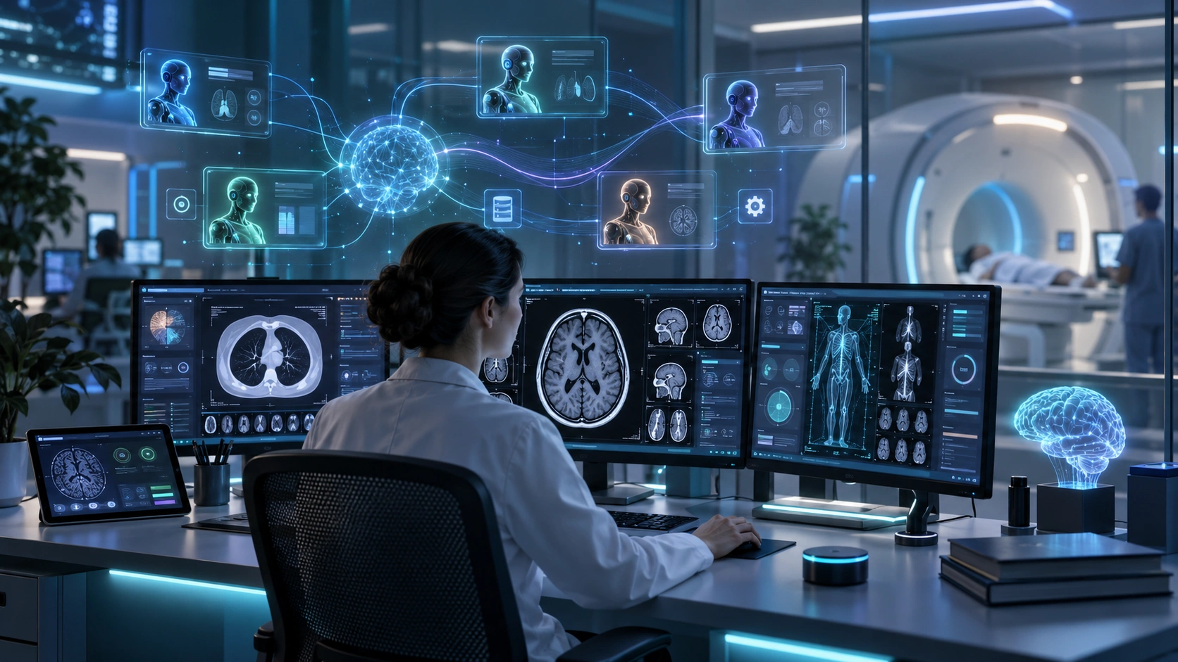
Earlier generations of medical imaging AI were designed for individual tasks, such as detecting a lung nodule, identifying a suspected brain bleed, or flagging a pneumothorax. Today’s emerging systems go further. Agentic AI in radiology can coordinate multiple specialized AI agents across an entire workflow—from worklist triage and prior-study retrieval to image analysis, report drafting, and quality checking.
In July 2026, Harrison.Rad 1.5 became the first model reported in the supplied benchmark to pass the FRCR 2B Short Case examination, scoring 86.5% against a 73.2% pass mark. GPT-5.4 and Gemini 3 scored 44% and 38%, respectively, in the same benchmark and did not pass.
The important shift is therefore not simply “AI reads scans.” It is the move from isolated AI detection tools toward multi-agent medical imaging workflows that can coordinate several steps before a radiologist reviews and signs the final report.
Key Takeaways
- Radiology AI is evolving from single-purpose detection toward connected, multi-agent workflows.
- AI agents can assist with triage, prior-study retrieval, image analysis, measurements, reporting, and quality assurance.
- Domain-specific radiology foundation models can outperform general-purpose models on specialized clinical benchmarks.
- AI-assisted workflows can reduce reporting turnaround time and increase the number of studies radiologists can handle.
- AI-generated reports still require appropriate validation, safety controls, and radiologist oversight.
From Single Detectors to Agentic Radiology AI
Traditional radiology AI typically performs one defined task.
For example, an AI model may analyze a chest X-ray and flag a possible pneumothorax. Another system might examine a head CT for suspected intracranial haemorrhage. These tools can be valuable, but they generally operate as individual components of the broader workflow.
Agentic AI radiology changes the architecture.
Instead of asking one model to complete everything, a multi-agent system assigns different responsibilities to specialized agents. One agent can prioritize the worklist, another can retrieve previous examinations, another can analyze the images, and another can draft or review the report.
An orchestration layer coordinates these tasks.
A useful analogy is a hospital department where every team member has a specific responsibility. One person handles intake, another prioritizes urgent cases, a specialist examines the case, a medical secretary prepares documentation, and a senior professional reviews the final result.
The AI system follows a similar pattern—but at software speed.
Large language models and multimodal foundation models make this possible by allowing AI systems to reason about context, coordinate tasks, use tools, and connect outputs from different stages of the workflow.
What Is Agentic AI in Radiology?
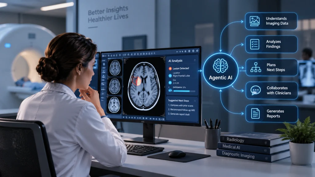
Agentic AI in radiology is a system of specialized AI agents coordinated to complete multiple connected steps of the medical imaging workflow. Rather than performing only image detection, agents can assist with worklist triage, prior-study retrieval, image analysis, measurements, report generation, quality checking, and communication. A radiologist remains responsible for clinical interpretation and final approval.
The goal is not simply to automate one task. It is to reduce repetitive work across the complete imaging journey.
The Radiologist Workflow: 9 Stages AI Is Automating
A typical radiology workflow contains several steps before a final report reaches the referring clinical team. Multi-agent AI can assist at each stage.
| Stage | Traditional Workflow | AI-Assisted Workflow |
| 1. Order Receipt | Radiologist reviews the imaging request and clinical context. | An NLP agent reads the request and flags missing information. |
| 2. Worklist Triage | Studies are manually prioritized by urgency. | A triage agent classifies cases and can reorder the worklist. |
| 3. Prior Study Retrieval | Radiologist searches PACS for relevant previous studies. | A retrieval agent finds and ranks relevant prior examinations. |
| 4. Image Analysis | Radiologist examines images for abnormalities. | A foundation model detects, segments, and localizes findings. |
| 5. Measurement & Quantification | Measurements are performed manually. | AI measures structures and tracks changes over time. |
| 6. Report Drafting | Radiologist dictates or writes the report. | A drafting agent generates a preliminary structured report. |
| 7. Quality Checking | Radiologist reviews their own report. | A QA agent checks the report against image findings. |
| 8. Critical Finding Notification | Referring clinicians may be contacted manually. | A communication agent can trigger alerts for critical findings. |
| 9. Report Delivery | Final report is signed and transmitted. | A workflow agent routes the signed report to connected systems. |
This illustrates why AI medical imaging workflow automation is becoming broader than simple image detection. Instead of replacing one activity, agentic systems can potentially reduce friction throughout the workflow.
How Multi-Agent Radiology AI Works
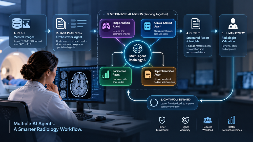
A multi-agent radiology system can be understood through five major components.
The Orchestrator Agent: The Coordinator
The orchestrator manages the other agents. It determines which task should happen first, delegates work, and handles exceptions.
Think of it as a senior radiologist coordinating a team of specialists. It does not necessarily perform every task itself; it decides who should perform each task and when.
The Triage Agent: The Emergency Nurse
The triage agent reviews incoming studies and categorizes them according to urgency.
For example, suspected stroke, intracranial haemorrhage, or pneumothorax can be prioritized so potentially critical cases reach the appropriate clinical team faster.
The supplied outline cites a 2026 study in which AI triage reduced CT-to-team notification time by 22 minutes in acute neurovascular imaging.
The Analysis Agent: The Specialist Reader
The analysis agent processes medical images through a vision foundation model. It can detect, segment, and localize abnormalities.
Models identified in the source document include Harrison.Rad 1.5, MedGemma, and MAIRA-2.
The outline also cites a meta-analysis reporting pooled sensitivity of 92% and specificity of 94% for intracranial haemorrhage detection across 73 studies.
The Drafting Agent: The Medical Secretary
Once image analysis is complete, a drafting agent can transform structured findings into a preliminary radiology report.
Instead of starting a report from an empty screen, the radiologist receives a draft that can be reviewed, edited, and signed.
The QA Agent: The Peer Reviewer
The QA agent provides another safety layer. It compares the image findings with the generated report and looks for inconsistencies, missing information, or potential errors.
This is especially important because even a low hallucination rate can have serious consequences in clinical reporting.
Foundation Models Powering Radiology AI
Foundation models provide the intelligence behind many emerging medical imaging LLM pipelines.
The supplied 2026 comparison highlights several specialized and general-purpose models:
| Model | Organization | Primary Strength | 2026 FRCR Score |
| Harrison.Rad 1.5 | Harrison.ai | VQA, report drafting, clinical exams | 86.5% — Passed |
| MedGemma 27B | Medical Q&A and chest X-ray | 18% — Failed | |
| Med-Gemini | Google DeepMind | Multimodal medical tasks | 38% — Failed |
| MAIRA-2 | Microsoft Research | Grounded chest X-ray reporting | Not evaluated |
| CheXagent 8B | Stanford | Chest X-ray interpretation | Below pass mark |
| GPT-5.4 | OpenAI | General medical reasoning | 44% — Failed |
| Gemini 3 | Long-context clinical documents | 38% — Failed |
One important lesson from these results is that general-purpose AI does not automatically equal specialized medical performance.
The source document reports that Harrison.Rad 1.5, trained on 6 million diagnostic imaging studies, achieved the strongest result in the cited FRCR benchmark. This reinforces the importance of domain-specific training when building AI systems for clinical environments.
Real-World Radiology AI Use Cases
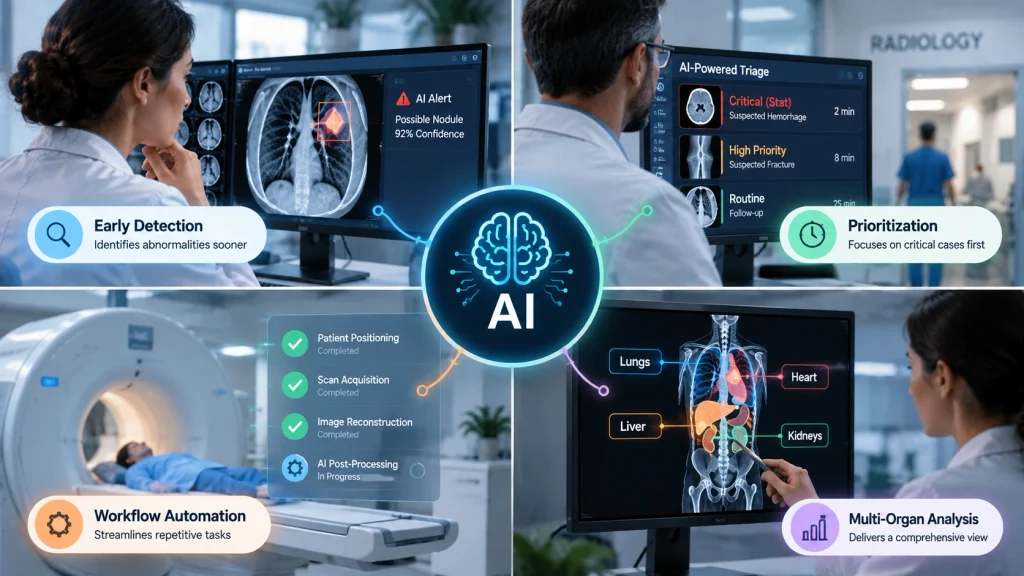
Emergency CT Triage
AI triage systems can analyze incoming head CT studies and flag suspected intracranial haemorrhage or other urgent findings. In the cited multi-centre study involving 3,409 brain CT scans, AI-assisted radiologists achieved 98.91% sensitivity and 99.83% specificity for ICH detection.
Automated Chest X-Ray Report Drafting
A drafting agent can prepare a preliminary structured report before the radiologist opens the case. The radiologist then reviews and edits the output instead of creating the report entirely from scratch.
Lung Nodule Surveillance
Multi-agent systems can track lung nodules across multiple CT examinations, automatically measure changes, identify growth, and support follow-up recommendations based on relevant guidelines.
Mammography and Breast Screening
AI can act as an additional reader in screening workflows, identifying suspicious regions before radiologist review. This can be particularly valuable where radiology departments face workforce shortages.
Oncology Prior-Study Comparison
Cancer follow-up frequently requires comparison of current scans with examinations from months or years earlier. A retrieval agent can locate relevant priors, an analysis agent can compare measurements, and a drafting agent can organize the findings into a structured response.
How Accurate Is AI in Medical Imaging in 2026?
Accuracy depends heavily on the task, model, dataset, and whether AI operates independently or alongside a radiologist.
The source document reports the following figures:
| Metric | Reported Result |
| AI-assisted report turnaround improvement | 15–20% faster |
| Radiologist throughput improvement | 10–20% more scans |
| ICH sensitivity — AI alone | 95.91% |
| ICH sensitivity — AI + radiologist | 98.91% |
| ICH specificity — AI + radiologist | 99.83% |
| Harrison.Rad 1.5 FRCR 2B | 86.5% |
| Harrison.Rad 1.5 hallucination rate | 5.7% |
| GPT-5.4 FRCR score | 44% |
| MedGemma 27B FRCR score | 18% |
The central point is that AI performance should not be interpreted as a single universal accuracy number. Clinical performance varies by modality, pathology, population, workflow, and level of human oversight.
Challenges and Limitations
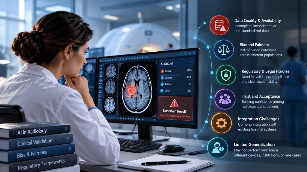
Despite the rapid development of radiology AI 2026, significant challenges remain.
Hallucination
AI-generated reports can contain information that is not supported by the underlying image. The supplied benchmark reports a 5.7% hallucination rate for Harrison.Rad 1.5, but no model has zero risk.
Regulation
There are more than 200 FDA-cleared AI medical devices, but many focus on specific detection or triage tasks. Broad, fully agentic systems that coordinate reporting workflows face additional regulatory and validation challenges.
Integration
Hospitals often depend on PACS, RIS, and EHR systems with fragmented workflows and proprietary interfaces. Connecting multiple AI agents to these environments can be technically complex.
Bias and Data Coverage
Foundation models may have uneven representation across modalities, populations, and geographic regions. Performance can vary when models encounter patient populations that differ from their training data.
Compute and Energy
Reasoning-intensive AI systems can require substantial computational resources. The source document notes energy-performance tradeoffs identified in SIIM 2026 research, making infrastructure costs an important consideration for large-scale deployment.
Is AI Replacing Radiologists?
AI is not simply replacing radiologists; it is increasingly being positioned as a workflow assistant that helps radiologists process more information and reduce repetitive tasks. The strongest model described in the supplied benchmark still operates within a workflow requiring human review and clinical judgment. AI can prioritize, analyze, draft, and check, while the radiologist remains responsible for interpreting the complete clinical context and approving the final report.
A useful analogy is a driver using an advanced navigation system. The system can make the journey faster and provide valuable information, but the driver remains responsible for decisions when unexpected situations occur.
The future of radiology is therefore more likely to be radiologist + AI rather than AI operating completely independently.
What Comes Next for Agentic Radiology AI?
The development of agentic AI radiology points toward increasingly connected imaging workflows.
Instead of using separate tools for detection, reporting, retrieval, and communication, hospitals may increasingly adopt systems where these capabilities work together through an orchestration layer.
The supplied roadmap suggests that future systems could move toward more autonomous reporting for routine studies and even agentic support for surgical planning.
For hospitals, the important question is therefore shifting from:
“Should we use radiology AI?”
to:
“Where can AI safely remove workflow bottlenecks while improving the radiologist’s ability to focus on clinical judgment?”
Key Takeaways
- Radiology AI is moving from isolated detection toward complete workflow assistance.
- Multi-agent systems divide complex tasks among specialized AI agents.
- Foundation models are becoming an important component of medical imaging AI.
- Human oversight remains essential because hallucinations, bias, integration, and regulatory issues persist.
- The most valuable AI systems may be those that improve the entire workflow rather than simply detecting one abnormality.
The next phase of medical imaging will not be defined only by how accurately AI can recognize an abnormality. It will be defined by how intelligently AI can coordinate the entire radiology workflow, from the moment an imaging order arrives to the moment a verified report reaches the clinical team.
Clinical Disclaimer: This blog is for informational purposes only and does not constitute medical advice. AI-generated reports require appropriate radiologist review and should not be treated as a substitute for qualified clinical judgment.